Hannah, a 22-year-old graduate student in Boston, started her first semester of a neuroscience PhD program in good health and ended it on a locked psychiatric ward, then a neurology floor, then a neurocritical care unit, with a diagnosis no one in her family had ever heard of. The first symptoms were flu-like and unremarkable: headache, low-grade fever, fatigue. Within two weeks she was paranoid, hearing voices, and convinced her advisor was poisoning her coffee. The on-call psychiatrist diagnosed first-episode psychosis and admitted her to an inpatient unit where she received olanzapine without improvement. By day five of admission she had a generalized tonic-clonic seizure, which prompted transfer to a teaching hospital where a sharp neurology fellow remembered the Susannah Cahalan story, ordered a lumbar puncture with NMDA receptor antibody testing, and discovered the autoimmune cause hiding behind what had looked like schizophrenia. A pelvic ultrasound revealed a small ovarian teratoma. Surgical removal, high-dose steroids, intravenous immunoglobulin, and eventually rituximab brought Hannah back, slowly, over fourteen months of recovery.
Hannah’s story is increasingly recognizable to neurologists and psychiatrists who follow the autoimmune encephalitis literature, but it remains routinely missed in community settings where anti NMDA encephalitis is not on the differential for new-onset psychosis. Anti-N-methyl-D-aspartate receptor encephalitis is an autoimmune disease in which antibodies target the GluN1 subunit of the NMDA receptor, producing a constellation of psychiatric, neurological, autonomic, and cognitive symptoms that evolves through recognizable stages. First described in 2007, the syndrome has become the most common autoimmune encephalitis in young adults and is a leading cause of identifiable encephalitis worldwide. Susannah Cahalan’s 2012 memoir Brain on Fire brought public attention to the disease and to the diagnostic odyssey many patients still endure.
Anti-NMDAR encephalitis as the great mental-illness mimic
Among autoimmune brain diseases, anti-NMDAR encephalitis stands out for how convincingly it impersonates primary psychiatric illness in its early stages. Patients typically present with anxiety, insomnia, mood lability, paranoia, and hallucinations that prompt psychiatric admission long before neurological signs make the autoimmune nature obvious. Clinicians who follow the literature now know to consider autoimmune encephalitis in any young patient with rapid-onset psychosis, particularly when accompanied by movement abnormalities, autonomic instability, seizures, or atypical features that do not fit cleanly into schizophrenia or bipolar disorder. The lesson from cases like Hannah’s is that first-episode psychosis evaluation must now include consideration of autoimmune causes, even when the presentation looks classical, especially in young women, in whom paraneoplastic forms tied to ovarian teratoma are most common.
The Susannah Cahalan story and public awareness
Susannah Cahalan was a 24-year-old reporter at the New York Post when she developed paranoia, hallucinations, seizures, and catatonia over several weeks in 2009. She cycled through psychiatric admissions and was on the verge of long-term institutionalization when neurologist Souhel Najjar performed a clock-drawing test that revealed dramatic hemispatial neglect, ordered the right antibody panel, and discovered the autoimmune cause. Her recovery story, told in Brain on Fire, has been credited with saving lives by educating clinicians and families about the disease. Public awareness now drives many of the parents and partners who present to emergency departments insisting their loved one’s psychiatric crisis be evaluated for autoimmune causes, and that advocacy has shortened the diagnostic delay considerably in well-resourced settings. Catatonia is a frequent feature of advanced anti-NMDAR encephalitis and demands the same diagnostic vigilance.

The four-stage progression
Anti-NMDAR encephalitis classically progresses through four overlapping stages. The prodromal stage involves nonspecific viral-like symptoms (headache, fever, fatigue, upper respiratory symptoms) lasting days to weeks. The psychiatric stage brings anxiety, insomnia, mood symptoms, paranoia, and hallucinations, often prompting psychiatric admission and antipsychotic treatment that frequently produces severe extrapyramidal side effects because of NMDA receptor dysfunction. The neurological stage adds seizures (focal or generalized), abnormal movements (orofacial dyskinesia, choreoathetosis, dystonic posturing), language disturbance, and decreased consciousness; autonomic instability with blood pressure swings, tachycardia, hypoventilation, and hyperthermia frequently emerges and may require ICU care. The recovery stage extends over months to years and involves slow improvement in cognition, behavior, and motor function, with persistent deficits in memory, executive function, and emotional regulation in many survivors.
CSF testing and the diagnostic workup
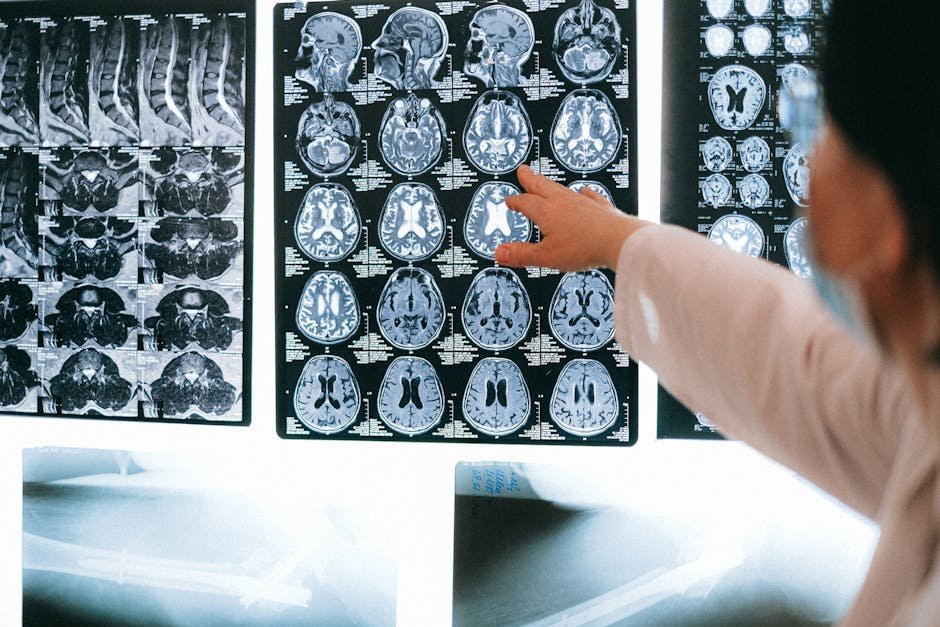
Definitive diagnosis requires detection of NMDA receptor antibodies in cerebrospinal fluid, which is more sensitive and specific than serum testing alone. CSF studies typically also show lymphocytic pleocytosis (mild to moderate elevation in white blood cells) and oligoclonal bands. MRI is normal in approximately half of cases but may show medial temporal lobe abnormalities or transient cortical and cerebellar enhancement. EEG frequently shows generalized slowing, focal abnormalities, or the characteristic extreme delta brush pattern in severely affected patients. Tumor screening is essential because of the strong paraneoplastic association: pelvic ultrasound or MRI in women to evaluate for ovarian teratoma, testicular ultrasound in men, and computed tomography of the chest, abdomen, and pelvis when initial screening is negative but suspicion remains high.
The ovarian teratoma association
In women between roughly 15 and 45 years of age, ovarian teratoma is identified in approximately half of anti-NMDAR encephalitis cases, and tumor removal is a critical component of treatment. The teratoma contains nervous tissue that expresses NMDA receptors, triggering the autoimmune response that crosses into the central nervous system. Tumors may be small enough to require detailed imaging or even bilateral oophorectomy in cases of pathologically definite paraneoplastic disease without identifiable mass. Tumor removal alone produces partial improvement, but combined immunotherapy is required for full recovery. In patients without identified tumors, the disease is presumed idiopathic, and recurrence rates are higher than in tumor-positive patients who undergo definitive resection. The National Institute of Neurological Disorders and Stroke at ninds.nih.gov maintains current research summaries on autoimmune encephalitis.
Treatment: immunotherapy in stepwise escalation
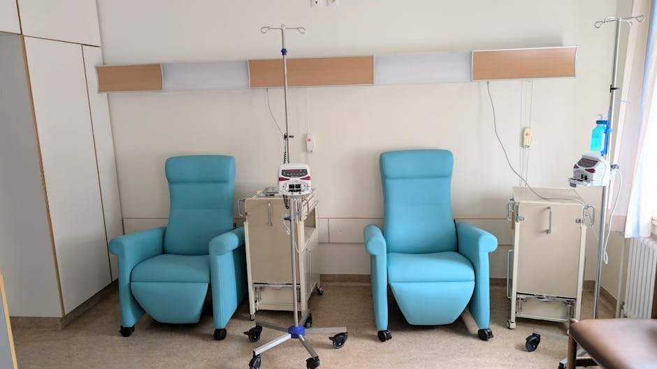
First-line immunotherapy combines high-dose intravenous methylprednisolone (typically 1 gram daily for five days), intravenous immunoglobulin (2 grams per kilogram divided over five days), and plasmapheresis or therapeutic plasma exchange. These therapies are often given concurrently in severe cases. Second-line therapy with rituximab and cyclophosphamide is added when first-line response is incomplete, which is common in tumor-negative cases and in patients with delayed diagnosis. Tumor resection when applicable is performed alongside immunotherapy. Maintenance immunosuppression with mycophenolate or azathioprine may be continued for one to two years to prevent relapse, particularly in tumor-negative patients. Symptomatic management includes seizure control, autonomic stabilization, careful selection of antipsychotic agents (avoiding high-potency dopamine antagonists when possible because of dyskinesia risk), and intensive rehabilitation.
- First-line: high-dose corticosteroids, IVIG, plasmapheresis, tumor resection if applicable
- Second-line: rituximab and cyclophosphamide for refractory cases
- Maintenance: mycophenolate mofetil or azathioprine for one to two years post-recovery
- Emerging therapies: bortezomib, tocilizumab in case series for treatment-resistant disease
- Symptomatic: anticonvulsants, autonomic stabilization, ICU support during peak severity
- Rehabilitation: physical, occupational, speech, and cognitive therapy during recovery
The recovery timeline
Recovery from anti-NMDAR encephalitis is typically slow and incomplete. With prompt treatment, approximately 75 to 80 percent of patients experience substantial recovery over twelve to twenty-four months, although residual deficits in memory, attention, executive function, and emotional regulation are common. Return to work or school often requires significant accommodations, and many survivors describe the cognitive recovery as lasting two to four years. Relapses occur in approximately 12 to 25 percent of patients and require renewed immunotherapy. Mortality is approximately 5 to 7 percent in the modern era of recognized disease and aggressive treatment, down from much higher rates before the syndrome was characterized. Earlier diagnosis correlates strongly with better outcomes, which is why awareness among psychiatric providers matters so much. Schizophrenia treatment programs increasingly include autoimmune encephalitis screening protocols for new-onset psychosis cases.
Finding autoimmune encephalitis specialists
Diagnostic and treatment expertise is concentrated at academic medical centers with neuroimmunology programs. The Autoimmune Encephalitis Alliance maintains a directory of specialist clinicians and centers of excellence across the United States. Major referral centers include Massachusetts General Hospital, the University of Pennsylvania, Mayo Clinic, Cedars-Sinai, and several university hospitals on the West Coast. For families navigating an active diagnosis, requesting transfer to such a center early in the hospital course often shortens diagnostic delay and improves treatment optimization. Telemedicine consultation has become more available since 2020 and can connect community hospitals to neuroimmunology expertise during initial workup. Family advocacy is frequently the deciding factor in obtaining the right antibody panel and the right treatment escalation in a timely manner.
Frequently asked questions about anti-NMDAR encephalitis
How is anti-NMDAR encephalitis different from schizophrenia?
Schizophrenia rarely produces seizures, autonomic instability, or movement abnormalities, while anti-NMDAR encephalitis typically does. CSF analysis with antibody testing reliably distinguishes the two when performed.
Can men get anti-NMDAR encephalitis?
Yes, although women account for the majority of cases. Male patients more often have idiopathic disease without identifiable tumor and may have testicular germ cell tumors as paraneoplastic triggers.
Will I fully recover?
Most treated patients experience substantial recovery over one to two years, but cognitive and emotional residua are common. Earlier treatment is associated with better long-term outcomes.
Can it come back?
Relapse occurs in 12 to 25 percent of cases, more often in tumor-negative patients. Maintenance immunosuppression reduces relapse risk, and ongoing follow-up with a neuroimmunologist is recommended.
Should children with new psychosis be tested?
Yes. Pediatric anti-NMDAR encephalitis exists, often presenting with behavioral changes, sleep disturbance, and movement abnormalities before psychotic features. CSF antibody testing is part of the standard pediatric workup for new-onset psychosis.
The bottom line
Anti-NMDAR encephalitis is a treatable autoimmune brain disease that mimics primary psychiatric illness early in its course and can produce excellent recovery when recognized and treated promptly. Any young patient presenting with new-onset psychosis accompanied by seizures, movement abnormalities, autonomic instability, or atypical features should be evaluated with cerebrospinal fluid antibody testing and tumor screening. Family advocacy and clinician awareness shorten the diagnostic odyssey and improve outcomes. The National Institutes of Health at nih.gov hosts research summaries and patient resources on autoimmune encephalitis.
If you are in crisis or experiencing thoughts of suicide, call or text 988 to reach the Suicide and Crisis Lifeline, available twenty-four hours a day across the United States.
This article is for educational purposes only and does not constitute medical advice. Always consult a qualified neurologist, psychiatrist, or autoimmune encephalitis specialist regarding diagnosis and treatment of suspected autoimmune brain disease.